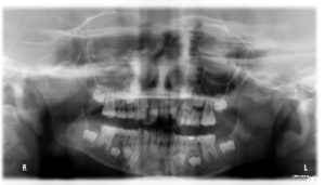
Dental examinations include the following:
-
- Digital x-rays to assess proper tooth development and healthy eruption of primary and permanent teeth
- Thorough examination of the teeth, looking for areas of decay, teeth that will be exfoliating (coming out soon), future orthodontic needs, and any treatment diagnosis
- Head, neck, and mouth visual examination
The digital dental radiographs are taken at least once per year for children at low risk for dental caries and every 6 months for children at high risk. They are only used as needed for diagnosis. If a visual examination revealed spacing between the teeth allow for complete visual view of all sides of the teeth, then the dentist may opt not to take any radiographs.
 Dental radiographs serve as an adjunct to the dentist’s in best diagnosis and treatment. The American Dental Association (ADA) encourages dentists and patients to discuss dental treatment recommendations, including the need for X-rays, to make an informed decision together. Radiation exposure associated with dentistry represents a minor contribution to the total exposure from all sources, including natural and man-made.
The National Council on Radiation Protection and Measurements (NCRP) has estimated that the mean effective radiation dose from all sources in the U.S. is 6.2 millisieverts (mSv) per year, with about half of this dose (i.e., 3.1 mSv) from natural sources (e.g., soil, radon) and about 3.1 mSv from man-made sources. About half of the man-made radiation exposure is related to CT scanning. Dental radiographs account for approximately 2.5 percent of the effective dose received from medical radiographs and fluoroscopies. While radiation exposure is low with digital radiographs, no one should receive more radiation than absolutely necessary. Protective lead aprons and thyroid collars should be used, especially for pregnant women, women of childbearing years and children.
