Categories:
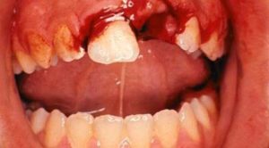
 Most injuries to primary teeth occur at 18 – 30 months of age, the toddler stage. As they learn to walk, they may fall forward landing on their hand and knees. The teeth most frequently injured are the upper central incisors, especially with protruding teeth being two to three times more prone. There are various types of injuries:
Luxation: This is the displacement of teeth with damage to the supporting structures which include periodontal ligament (PDL) and alveolar bone. The PDL supports the tooth to the socket.
- Concussion: The tooth is not mobile and is not displaced
- Mobility: The tooth is loosened but is not displaced from its socket
- Intrusion: The tooth is driven into its socket. This compressed PDL commonly causes a crushing fracture of the alveolar socket.
- Extrusion: This is the dislocation or displacement of the tooth from the socket. The PDL is torn.
- Lateral Extrusion: The tooth is displaced in a labial, lingual or lateral direction. The PDL is usually torn and fracture of the supporting alveolar bone.
- Avulsion: The tooth is completely displaced from the alveolus. The PDL is severed and fracture of the alveolus may occur.
The sequel to traumatized teeth may be devitalization due to lack of blood supply to the teeth. The potential sequel includes:
- Pulp hyperemia: Sensitive to percussion, transillumination of the crown with bright light. The condition may be reversible.
- Pulp Hemorrhage: Due to hyperemia there is bleeding from the capillaries, leaving blood pigments deposited in the dentinal tubules. In mild cases, the blood is resorbed and very little discoloration or may be lighter in color in a few weeks. In severe case, the discoloration continues for the life of the tooth.
- Calcific Metamorphosis: The pulp chamber and canal are gradually obliterated by progressive deposition of dentin. These teeth usually appear yellow and no treatment is recommended.
- Pulp Necrosis: In the absence of blood circulation, the pulp becomes necrotic (dead). Radiograph may show radiolucency indicating a granuloma or a cyst with often a parulis clinical evident at the level of the tooth’s root apex. The treatment required may be a pulpectomy with a resorbable material (zinc oxide and eugenol) or extraction due to risk to the permanent development teeth.
- Inflammatory Resorption: This can occur either on the outside root surface or inside the pulp chamber or canal. If treated, the tooth is treated with resorbable zinc oxide paste.
- Replacement Resorption: Also termed ankylosis, results after irreversible injury to the PDL. Alveolar bone directly contacts and becomes fused with the root surface. If there is delayed or ectopic the eruption of the permanent tooth then extraction should be performed.
